The retinal physicians at Plessen Ophthalmology have received advanced subspecialty training in the diagnosis and management of diseases of the retina, vitreous, and macula. Their clinical expertise and surgical experience uniquely positions Plessen Ophthalmology to handle a variety of diseases of the retina on St. Croix and St. Thomas. For more information about the conditions we treat and how these conditions are treated, please call or visit one of our locations.
Retinal diseases vary widely, but most of them cause visual symptoms. Retinal diseases can affect any part of your retina, a thin layer of tissue on the inside back wall of your eye. The retina contains millions of light-sensitive cells (rods and cones) and other nerve cells that receive and organize visual information. Your retina sends this information to your brain through your optic nerve, enabling you to see.
Treatment is available for some retinal diseases. Depending on your condition, treatment goals may be to stop or slow the disease and preserve, improve or restore your vision. Untreated, some retinal diseases can cause severe vision loss or blindness.
Symptoms:
Many retinal diseases share some common signs and symptoms. These may include:
- Seeing floating specks or cobwebs
- Blurred or distorted (straight lines look wavy) vision
- Defects in the side vision
- Lost vision
You may need to try looking with each eye alone to notice these.
Conditions We Treat:
- Diabetic Eye Screenings
- Diabetic Retinopathy
- Age-Related Macular Degeneration (Dry and Wet)
- Macular Hole
- Epiretinal Membrane
- Retinal Detachment
- Retinal Vein Occlusion and Artery Occlusion
- Hereditary Retinal Diseases
- Uveitis
- Intraocular Tumors
- Cataract Surgery Complications
- Trauma
- Proliferative Vitreoretinopathy
Diagnostic Services:
- Optical Coherence Tomography (Cirrus OCT)
- Fluorescein Angiography (FA)
- Ocular Ultrasound (B-Scan)
- Indocyanine Green (ICG)
- Ultra-wide Field Fundus Photography and FA
- Visual Field Testing
- Diopsys Electroretinography and Visual Evoked Potential Testing
Therapeutic Services:
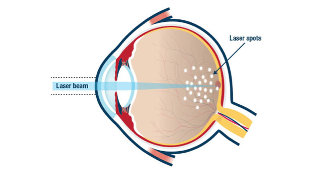
Laser Photocoagulation –
Laser photocoagulation is a controlled method of delivering focused laser power to the retina, creating limited burns. Laser photocoagulation has been used in retinal surgery for over many years. Laser photocoagulation is used in the office to treat a variety of conditions including: Diabetic macular edema, proliferative diabetic retinopathy, retinal vein occlusion, retinal tears or holes, retinal detachment, high risk lattice degeneration, and retinal arterial microaneurysm. Depending on the condition being treated, laser may be delivered with the surgeon wearing a headset (indirect ophthalmoscope) while the patient lays in an examination chair, or with the surgeon and patient sitting on opposite sides of table-mounted equipment (slit lamp).
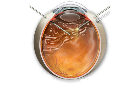
Vitrectomy –
Vitrectomy is the surgical removal of the vitreous gel from the middle of the eye. This procedure may be done for several reasons. To remove scar tissue membranes from the retinal surface, to repair macular holes, to repair retinal detachments, to remove vitreous hemorrhage, as well as other less frequent indications. Patients with diabetes are particularly prone to retina problems for which a vitrectomy may be recommended (to remove blood in the vitreous gel caused by abnormal vessel growth and vessel hemorrhage). During a vitrectomy, the surgeon inserts small instruments into the eye, cuts the vitreous gel, and removes it by suction. After removing the vitreous gel, the surgeon may treat the retina with a laser (photocoagulation), cut or remove fibrous or scar tissue from the retina, flatten areas where the retina has become detached, or repair tears or holes in the retina or macula.At the end of the surgery, saline, air or a gas (perfluropropane), or silicone oil may be injected into the eye to replace the vitreous gel to restore normal pressure in the eye.
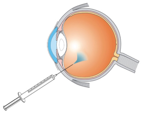
Intravitreal Injection –
Intravitreal injection is a method of delivering medication directly into the back part of the eye, the vitreous cavity. The retina lines the vitreous cavity, and medications delivered into the vitreous cavity can act directly on the retina and adjacent tissues. Intravitreal injections are given in the office using topical anesthesia (drops or gel placed on the surface of the eye). The different types of intravitreal injection medication offered at Plessen Ophthalmology include:
- Avastin
- Ozurdex
- Triessence/Kenalog
- Eylea
Most patients who receive an intravitreal injection will see floaters in their vision for an average of 2–10 days. These floating dark spots are from the medication actually drifting around inside your eye. Sleeping with your head slightly elevated can reduce floaters. They are harmless and expected. Mild swelling, irritation, itching and/or redness in and around the eye for a Day or Two, is not uncommon. These symptoms are usually reduced by using lubricating eye drops such as Tears or Refresh. On occasion, the treated eye can look blood-red following the injection. This is caused by a tiny leaking blood vessel in the “whites” of the eye. As long as this blood is not coming out of the eye, it is harmless and will slowly clear up in a Week to 10 days. Please use it as directed. You will be given a follow-up appointment to see your doctor at Plessen Ophthalmology. It is important to keep this appointment so your doctor can gauge the effectiveness of your treatment.
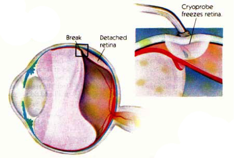
Cryotherapy –
Ocular cryotherapy is the therapeutic use of cold temperatures to treat disorders of the lids or eyes. Cryotherapy involves placing a very cold metal probe against the wall of the eye so that all of the eye’s layers are frozen, including the retina that lines the inside of the eye. Cryotherapy creates an adhesive scar that seals the retina against the wall of the eye. The effect of cryotherapy (the adhesive scar) is similar to laser treatment, but the treatment effect is accomplished using cold rather than heat. Unlike laser, cryotherapy can be used to form an adhesion that will hold detached retina to the eye wall once the retina attaches. In this way, cryotherapy is like glue where laser is more like a stapler. For laser to work, the retina has to be already attached. It takes cryotherapy about a week to form a strong adhesion.
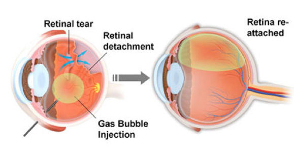
Pneumatic Retinopexy –
Pneumatic retinopexy (PR) is an office-based non- incisional procedure that has become a well-accepted alternative to scleral buckling and vitrectomy for the repair of selected cases of rhegmatogenous retinal detachment. If you have pneumatic retinopexy, your eye doctor will inject an expanding gas bubble into your eye. He or she will position you so that the bubble floats over the detached area and pushes it against the back of your eye. Your eye doctor then uses a freezing device to seal the retina against the wall of the eye.